Home

ablassen Rede Mut leading edge to leading edge echo Verfault Kessel Melodisch

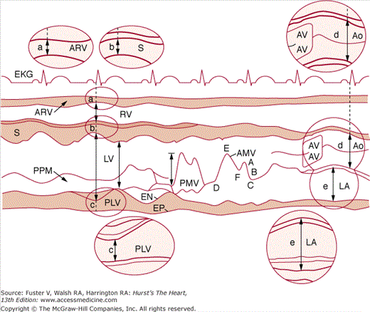
Comparison between M-mode border measurement conventions. The Standard... |  Download Scientific Diagram
Comparison between M-mode border measurement conventions. The Standard... | Download Scientific Diagram

Individual reference values for 2D echocardiographic measurements. The  Stockholm – Umeå Study - Svedenhag - 2015 - Clinical Physiology and  Functional Imaging - Wiley Online Library
Individual reference values for 2D echocardiographic measurements. The Stockholm – Umeå Study - Svedenhag - 2015 - Clinical Physiology and Functional Imaging - Wiley Online Library
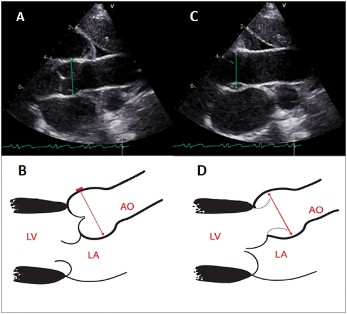
New Screening Tool for Aortic Root Dilation in Children with Marfan  Syndrome and Marfan-Like Disorders | SpringerLink
New Screening Tool for Aortic Root Dilation in Children with Marfan Syndrome and Marfan-Like Disorders | SpringerLink

EACVI survey on standardization of cardiac chambers quantification by  transthoracic echocardiography
EACVI survey on standardization of cardiac chambers quantification by transthoracic echocardiography

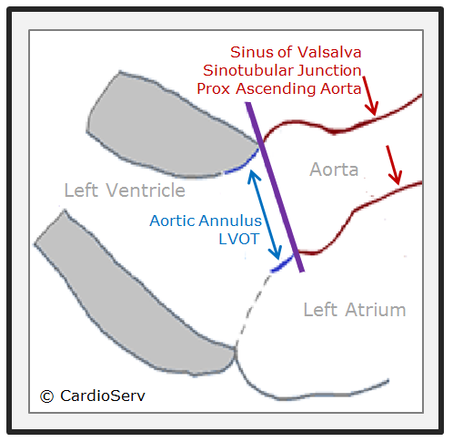
How to Master Aortic Measurements With These 5 Techniques

Standard method for ultrasound imaging of coronary artery in children -  Fuse - 2010 - Pediatrics International - Wiley Online Library
Standard method for ultrasound imaging of coronary artery in children - Fuse - 2010 - Pediatrics International - Wiley Online Library

頸動脈エコーの基礎>頸動脈壁の超音波像>超音波検査の計測原理 | 早期動脈硬化研究会

Figure 2 | The Methodology of Doppler-Derived Central Blood Flow  Measurements in Newborn Infants
Figure 2 | The Methodology of Doppler-Derived Central Blood Flow Measurements in Newborn Infants

Echocardiography | Thoracic Key

Supplemental Materials for Reference Values for Mid-Ascending Aorta  Diameters by Transthoracic Echocardiography in Adults - American Journal of  Cardiology
Supplemental Materials for Reference Values for Mid-Ascending Aorta Diameters by Transthoracic Echocardiography in Adults - American Journal of Cardiology

Comparison between M-mode border measurement conventions. The Standard... |  Download Scientific Diagram
Comparison between M-mode border measurement conventions. The Standard... | Download Scientific Diagram

How to Master Aortic Measurements With These 5 Techniques

The New Dimension in Aortic Measurements - Use of the Inner Edge  Measurement for the Thoracic Aorta in Australian Patients. | Semantic  Scholar
The New Dimension in Aortic Measurements - Use of the Inner Edge Measurement for the Thoracic Aorta in Australian Patients. | Semantic Scholar

The New Dimension in Aortic Measurements - Use of the Inner Edge  Measurement for the Thoracic Aorta in Australian Patients
The New Dimension in Aortic Measurements - Use of the Inner Edge Measurement for the Thoracic Aorta in Australian Patients

Aortic root measurement at sinus of Valsalva utilizing leading... |  Download Scientific Diagram
Aortic root measurement at sinus of Valsalva utilizing leading... | Download Scientific Diagram

THE AMERICAN SOCIETY OF ECHOCARDIOGRAPHY RECOMMENDATIONS FOR CARDIAC  CHAMBER QUANTIFICATION IN ADULTS: A QUICK REFERENCE GUIDE F
THE AMERICAN SOCIETY OF ECHOCARDIOGRAPHY RECOMMENDATIONS FOR CARDIAC CHAMBER QUANTIFICATION IN ADULTS: A QUICK REFERENCE GUIDE F

A schematic representation of three measurement methods used to... |  Download Scientific Diagram
A schematic representation of three measurement methods used to... | Download Scientific Diagram

Tuğba Kemaloğlu Öz, Assoc Prof on Twitter: "Which method do u prefer? Leading  edge vs inner edge? #echofirst https://t.co/LZDxcPMsUd" / Twitter
Tuğba Kemaloğlu Öz, Assoc Prof on Twitter: "Which method do u prefer? Leading edge vs inner edge? #echofirst https://t.co/LZDxcPMsUd" / Twitter

Small animal cardiac ultrasound measurements | IMV imaging

Reference Values of Aortic Root in Male and Female White Elite Athletes  According to Sport | Circulation: Cardiovascular Imaging
Reference Values of Aortic Root in Male and Female White Elite Athletes According to Sport | Circulation: Cardiovascular Imaging

Reproducibility of ECG-gated Ultrasound Diameter Assessment of Small  Abdominal Aortic Aneurysms - European Journal of Vascular and Endovascular  Surgery
Reproducibility of ECG-gated Ultrasound Diameter Assessment of Small Abdominal Aortic Aneurysms - European Journal of Vascular and Endovascular Surgery

V.L.Sorrell, MD (@universityofky) on Twitter: "#JADEL; #JADIL How do you  measure the Aortic root? @ASE360 states leading edge to leading edge  end-diastole. CCT/CMR prefers inner edge to inner edge mid-systole. When  possible,
V.L.Sorrell, MD (@universityofky) on Twitter: "#JADEL; #JADIL How do you measure the Aortic root? @ASE360 states leading edge to leading edge end-diastole. CCT/CMR prefers inner edge to inner edge mid-systole. When possible,

Focused Echocardiographic Examination in the Emergency Room and Critical  Care Units Ronald E. Cuyco, MD, FPCC. - ppt video online download
Focused Echocardiographic Examination in the Emergency Room and Critical Care Units Ronald E. Cuyco, MD, FPCC. - ppt video online download

Aortic arch dimension measured using the leading edge method; 1) aortic...  | Download Scientific Diagram
Aortic arch dimension measured using the leading edge method; 1) aortic... | Download Scientific Diagram

Manual of Echocardiography

Indian Academy of Echocardiography e-Newsletter

THE AMERICAN SOCIETY OF ECHOCARDIOGRAPHY RECOMMENDATIONS FOR CARDIAC  CHAMBER QUANTIFICATION IN ADULTS: A QUICK REFERENCE GUIDE F
THE AMERICAN SOCIETY OF ECHOCARDIOGRAPHY RECOMMENDATIONS FOR CARDIAC CHAMBER QUANTIFICATION IN ADULTS: A QUICK REFERENCE GUIDE F

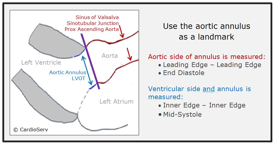
Aortic dimensions by multi-detector computed tomography vs. echocardiography  - Journal of Cardiology
Aortic dimensions by multi-detector computed tomography vs. echocardiography - Journal of Cardiology